It has been a hot summer in the Microscopy Labs here at RBGE as students and visitors from all over the world have been busy using our facilities. Here are just a few examples of the work that has been carried out as introduced by our guests….
Jia Dong
SEM Image of Elatostema rugosum
‘Hi! I’m Jia, an MSc student here from China. I’m using the Scanning Electron Microscope (SEM) to look at the floral development of the Nettle Family — Urticaceae.’
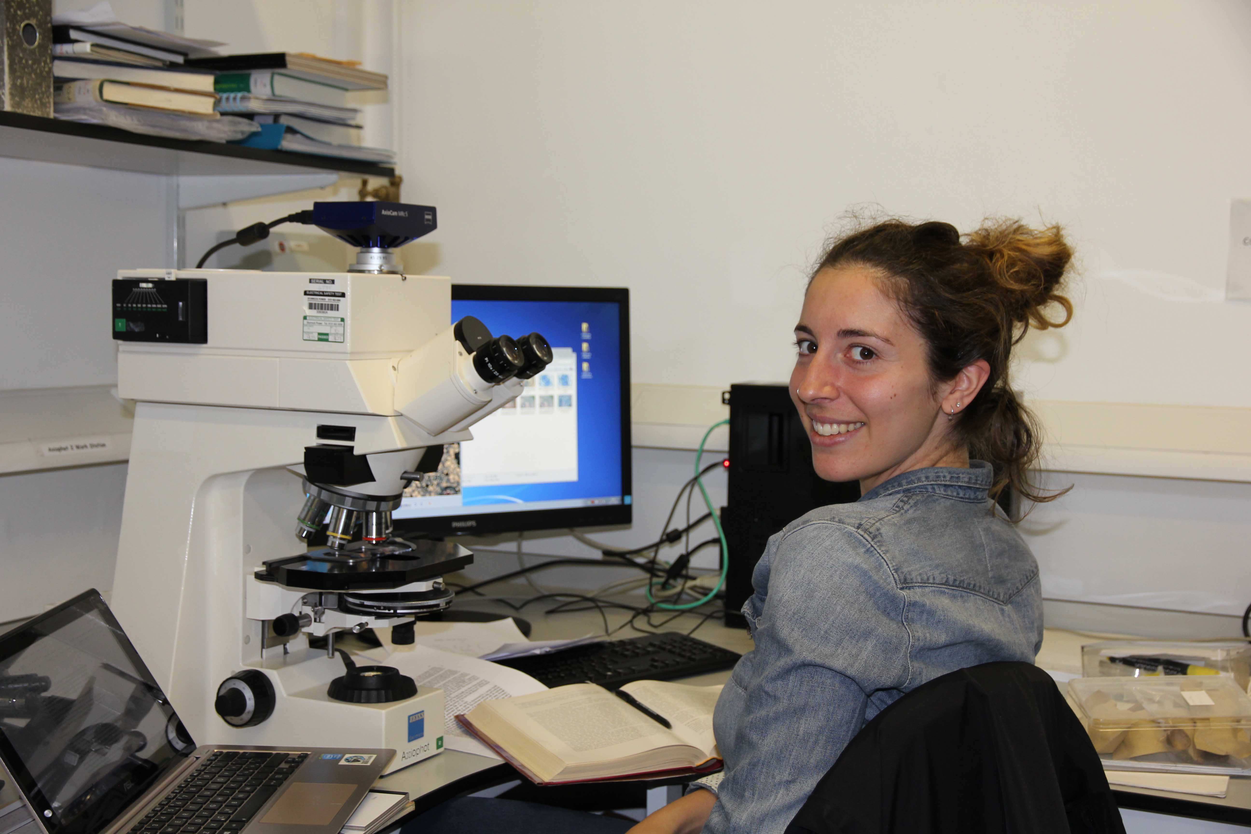
Ludovica Santilli
I am from Rome, Italy and I am using the light microscope to look at the fruits and seeds anatomy of Sabiaceae.
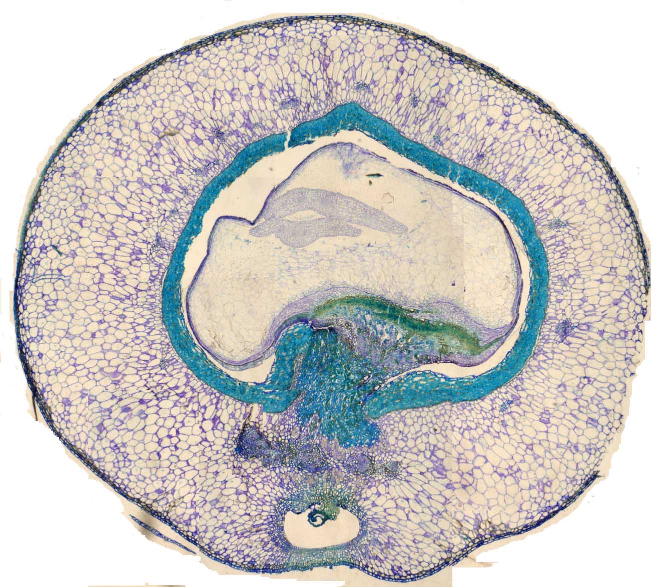
Stained Light Microscope Image of Meliosma cuneifolia
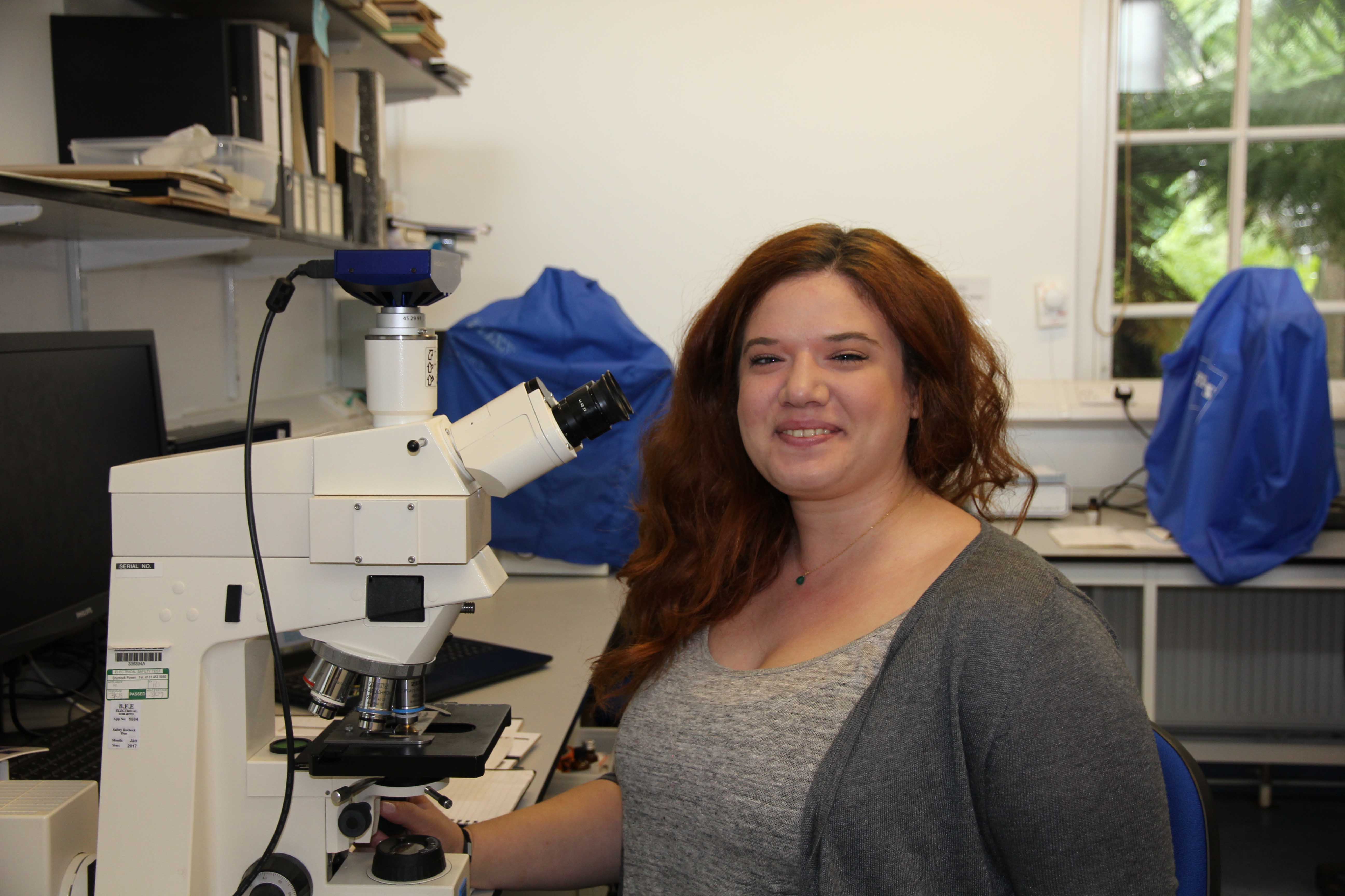
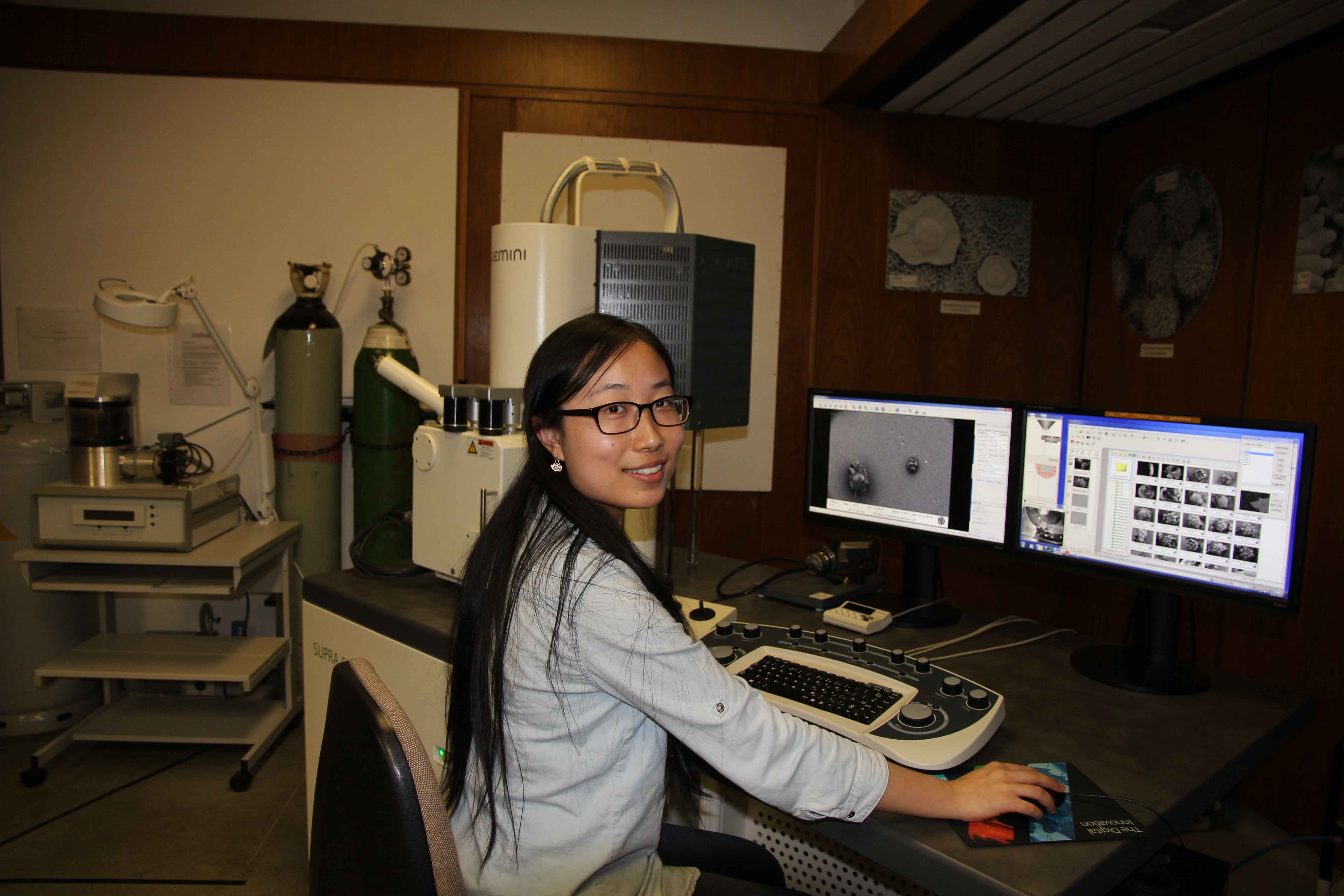
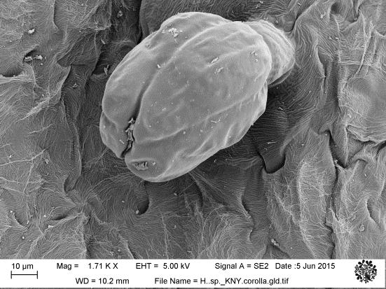
Marita Papagianni
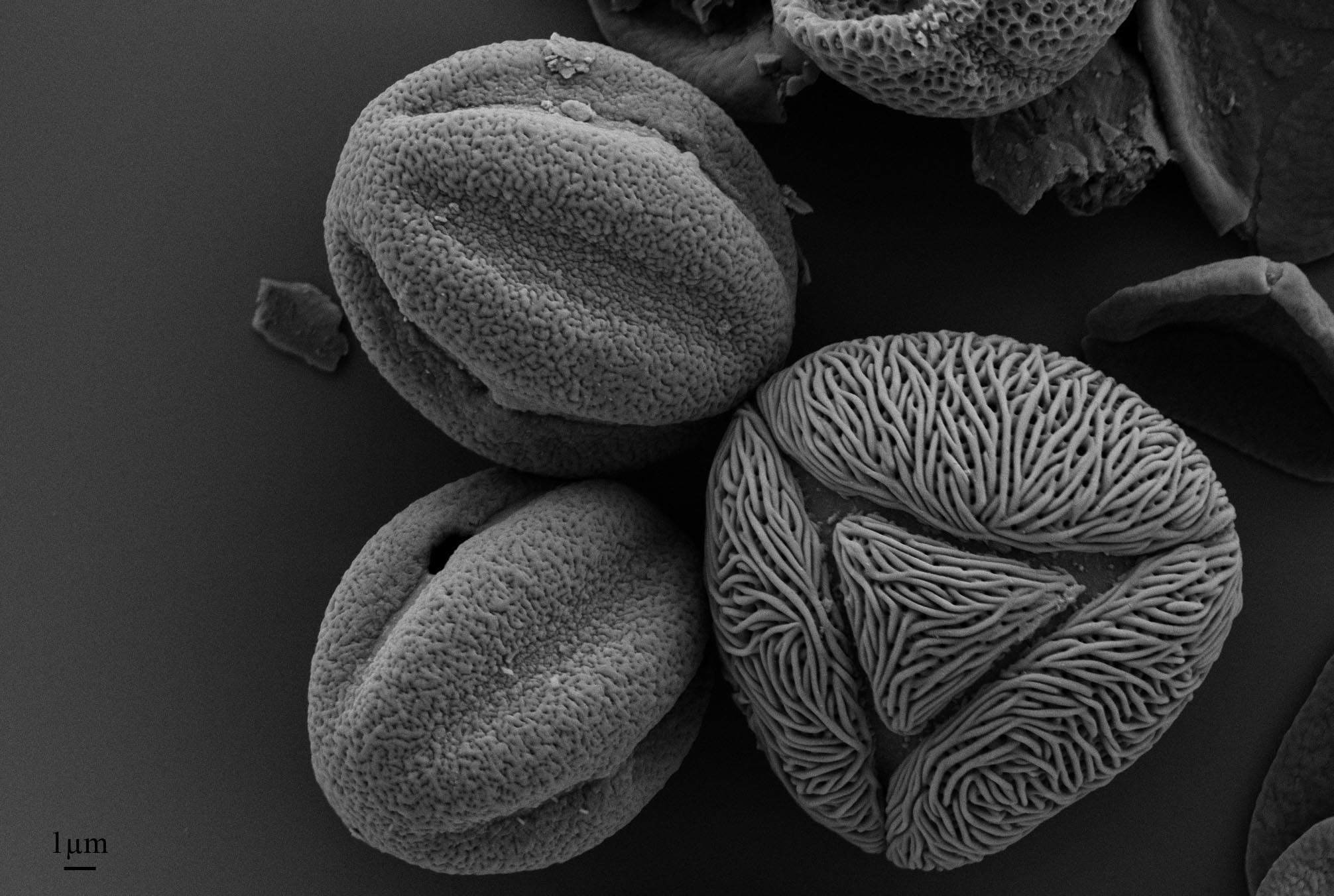
SEM of Pollen Extracted from Nepalese Honey
I am Marita Papagianni from Thessaloniki, Greece. I used SEM and light microscopy to photograph pollen grains extracted from Nepalese honey as a part of my MSc thesis. The pollen grains with the concavities on the left (SEM photo) belong in Melastomataceae and the triangular “noodle-like” pollen grain is from Schleichera oleosa (Sapindaceae).
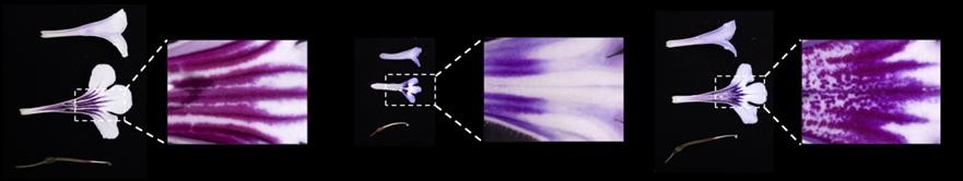
Light Microscope Images of Gesneriaceae Flowers
I am Yun-Yu Chen from Taiwan. I am using the Stemi stereo microscope to record the floral characters in Gesneriaceae species (with beautiful flowers!)

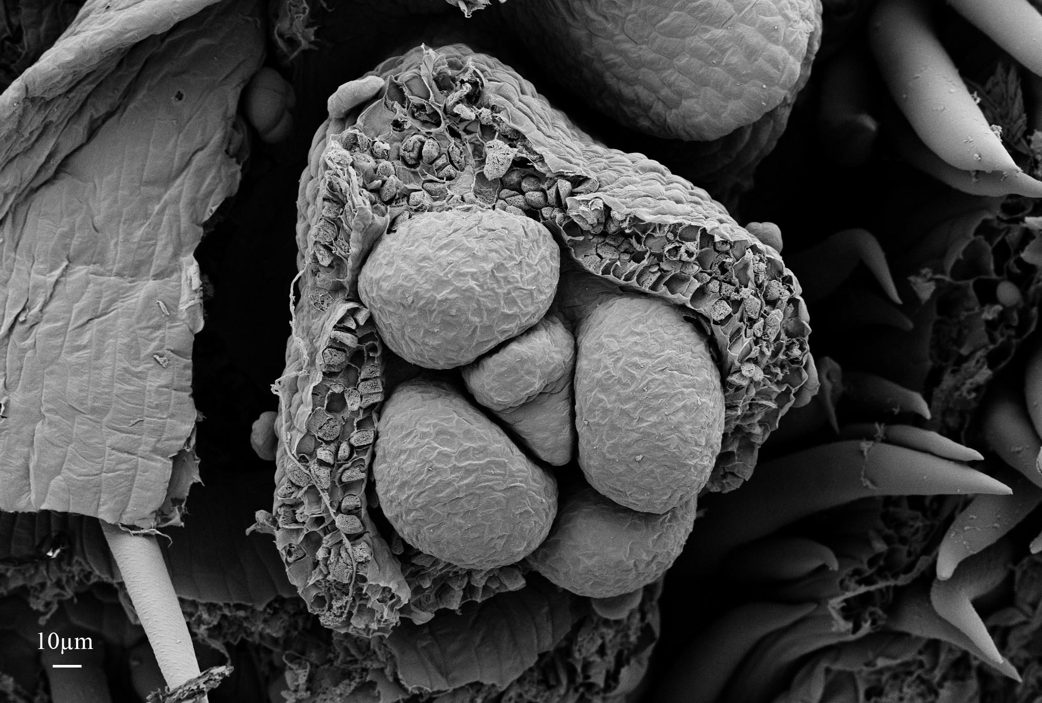
The ‘Caverns’ of Boletus calocystitides with its amazing cystidia (sterile cells on the Boletus tubes)
Hi all! I’m Serena Lee from the Singapore Botanic Gardens and for my summer project. I’m working on a sub group of Boletus, the Xerocomoids. I am simply loving the use of the SEM here as it brings me to worlds yet undiscovered . It feels as if I’m on a Mars land rover surveying the contours of the planet or in caverns, 20,000 leagues under the seas.
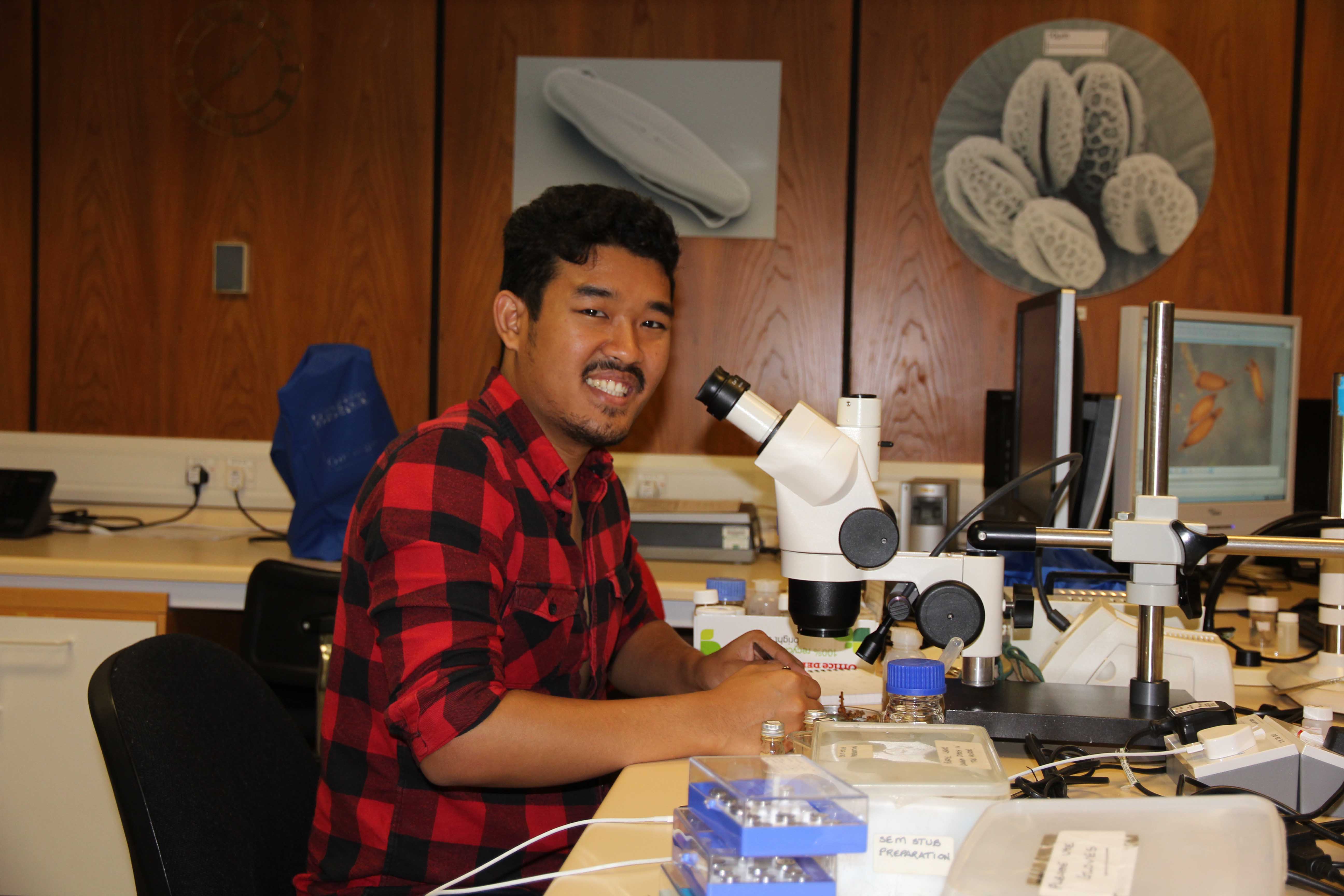
Pakkapol Thaowetsuwan
My name is Pakkapol Thaowetsuwan, a PhD student from Thailand. I am using SEM to investigate evolution and development of floral organs in Croton and related genera (Crotonoideae, Euphorbiaceae).
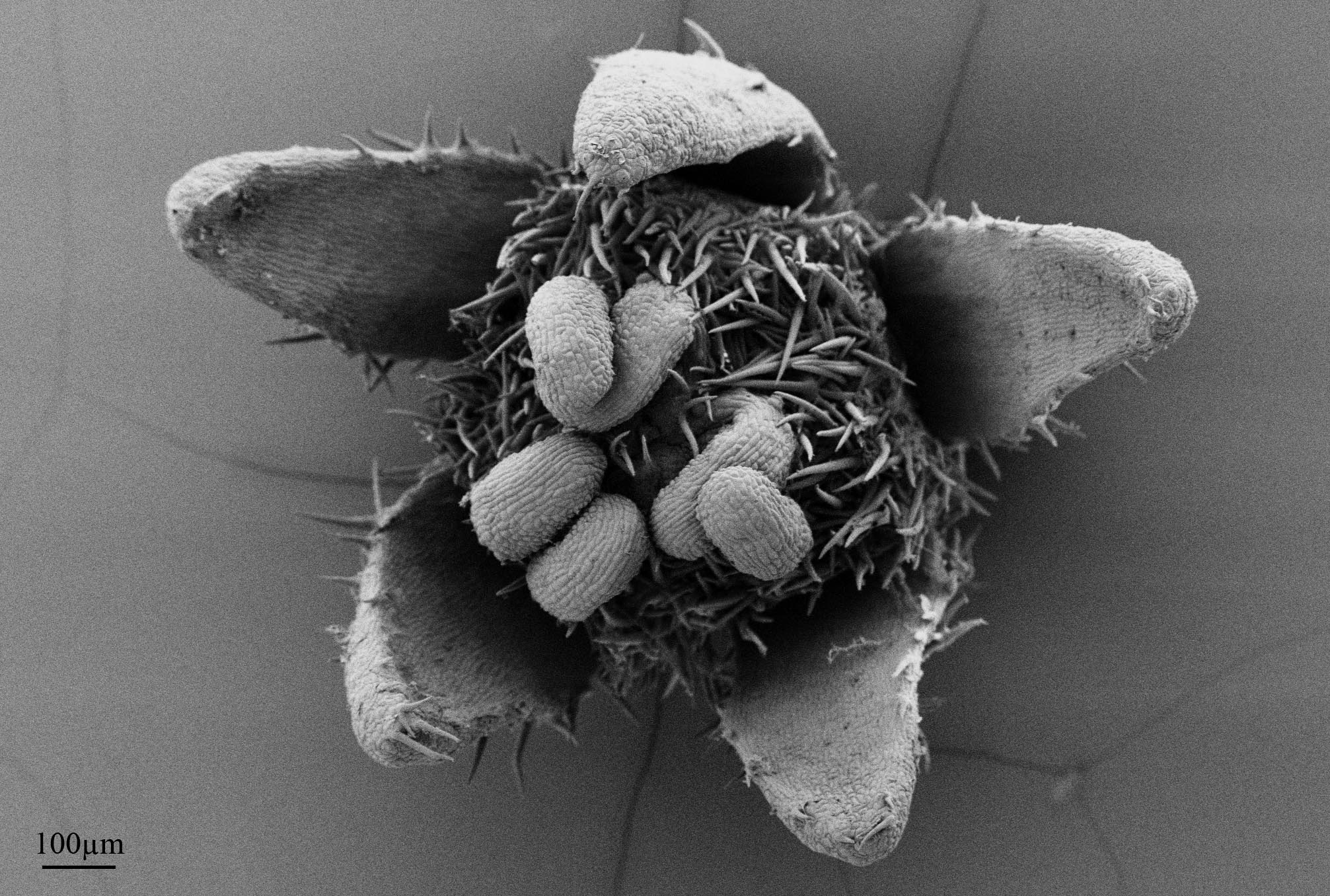
Croton trinitatis female flower
I am a Brazilian biologist, living in Switzerland and completing my MSc. studies in Botany and Ecology at the University of Basel under the supervision of Louis Ronse De Craene (Royal Botanic Garden of Edinburgh). I am very interested on Flower Morphology. An adaquate way of understanding flowers is by looking at how they originate. This is one of the main reasons why I am working with SEM. Felipe Toni
Felipe Toni
SEM Image of Sanguisorba officinalis

Subhani Ranasinghe
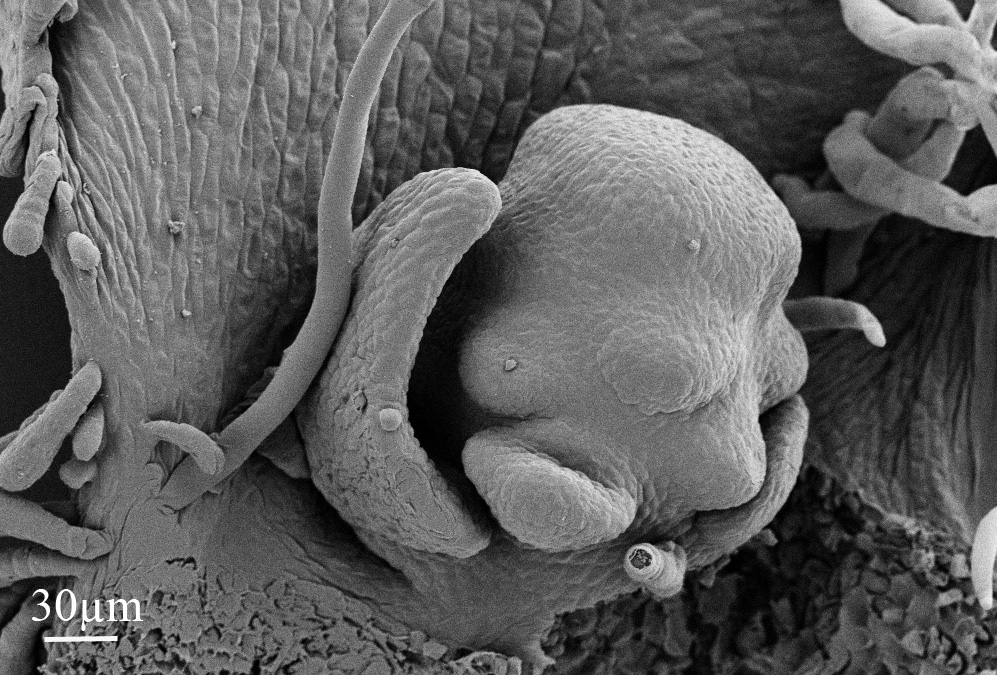
I am a third year PhD student from Sri Lanka working on “Taxonomy, molecular species delimitation, phylogeny and Biogeographic affinities of Sri Lankan Gesneriaceae”. I used the microscopic unit for studying of microscopic characters in particular, several indumentum characters, flowers and seeds using STEMI dissecting microscope and SEM. Subhani Ranasinghe
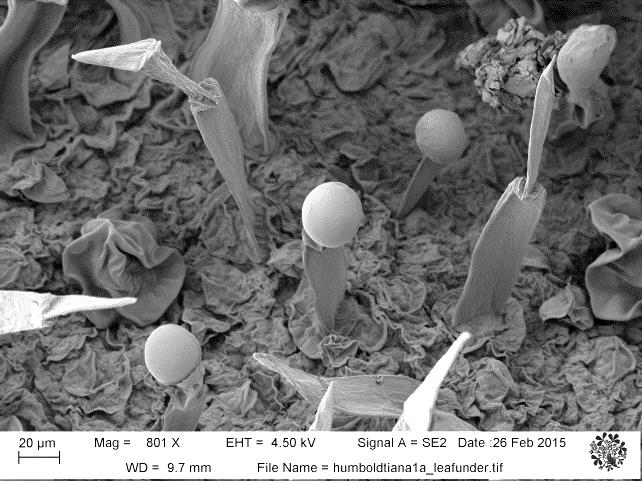
Different glandular structures of Henckelia humboldtiana (Gardner) A. Weber & B.L. Burtt under SEM
Anjum Farooqui
The pollen image of Schleichera is slightly different in ornamentation which I photographed from honey, western ghats India. would like to discuss more on this