After nearly 28 years at the Royal Botanic Garden Edinburgh (RBGE), Frieda Christie, our Microscopy Lab manager, will be retiring at the end of September.
To celebrate Frieda’s time with us, we’ve gathered some of our favourite Scanning Electron Microscope (SEM) images taken by Frieda since 2003.
Firstly, a little bit about Frieda….
Frieda started at RBGE in December 1993 as a Microscopist for the newly installed SEM, within the Department of Scientific and Technical Services. As the RBGE lab facilities have developed, so has Frieda’s role. She is now the Microscopy lab manager, in charge of all the research microscopes and the new(ish) SEM that was purchased in 2003. The investment in the SEM facilities at RBGE is an acknowledgment of the importance of having these facilities in-house in a taxonomic research institute.
In addition to the many scientific publications that Frieda has produced or contributed to, you can see many of Frieda’s images around the Gardens, on the walls of the John Hope Gateway, in the laboratory corridors or in some of our beautifully illustrated books. Frieda’s artistic flare has won her many awards including 1st Prize in the ‘Science as Art’ category in the Novartis/Daily Telegraph Images in Science Competition. In 2000 Frieda was awarded full sponsorship for her ‘Microscopic Artist’ Exhibition which toured several locations including the Edinburgh and Benmore Gardens, Dundee College of Art and finished its run at the Kelvingrove Gallery in Glasgow. And in 2020 her image of Rhododendron grande was chosen for the front cover of infocus magazine of the Royal Microscopical Society.
Coloured SEM image of an anatropous ovules of the Gesneriaceae Anna submontana
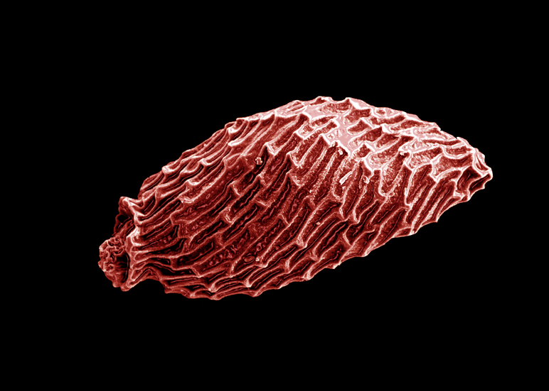
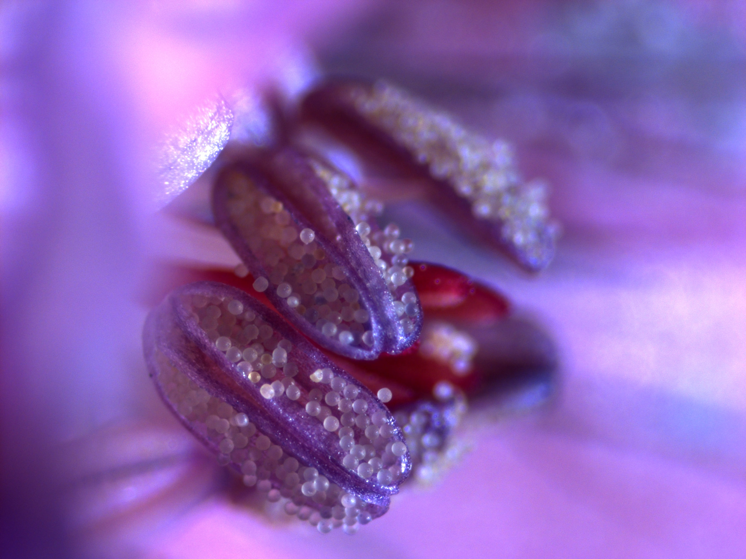
Coloured SEM image of pollen from Victoria ‘Longwood Hybrid’ (giant water lily) 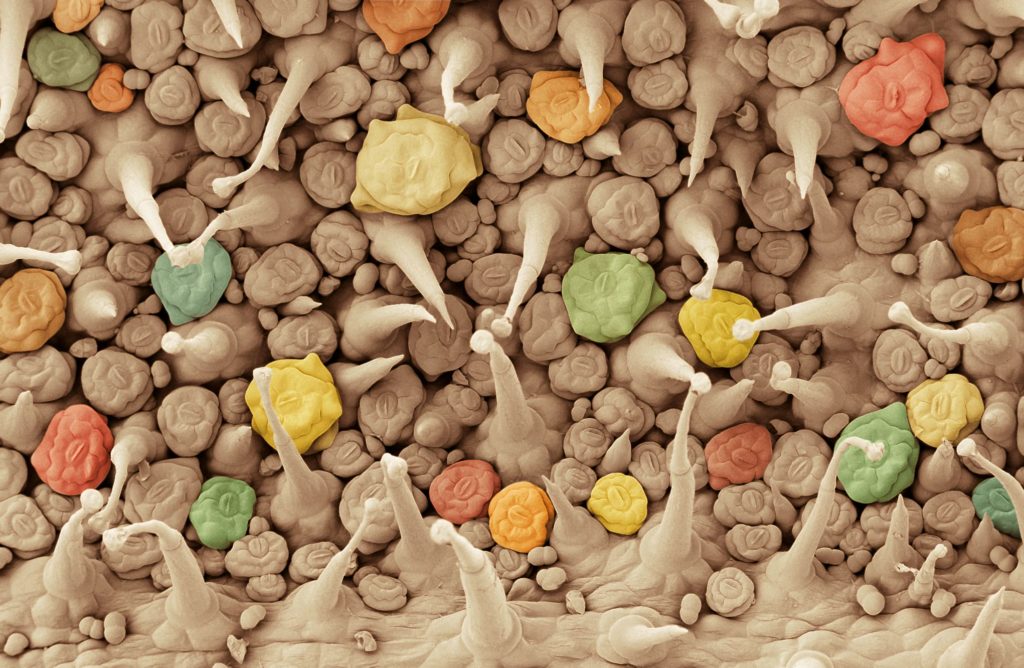
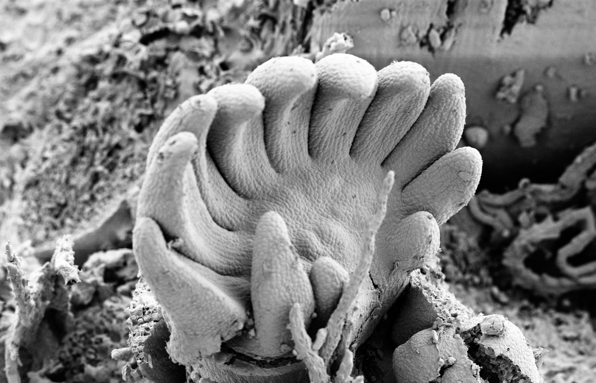
Coloured SEM image of the abaxial epidermal leaf surface of Streptocarpus suffruticosus (Gesneriaceae) with glandular and eglandular trichomes and stomatal turrets of varying size. 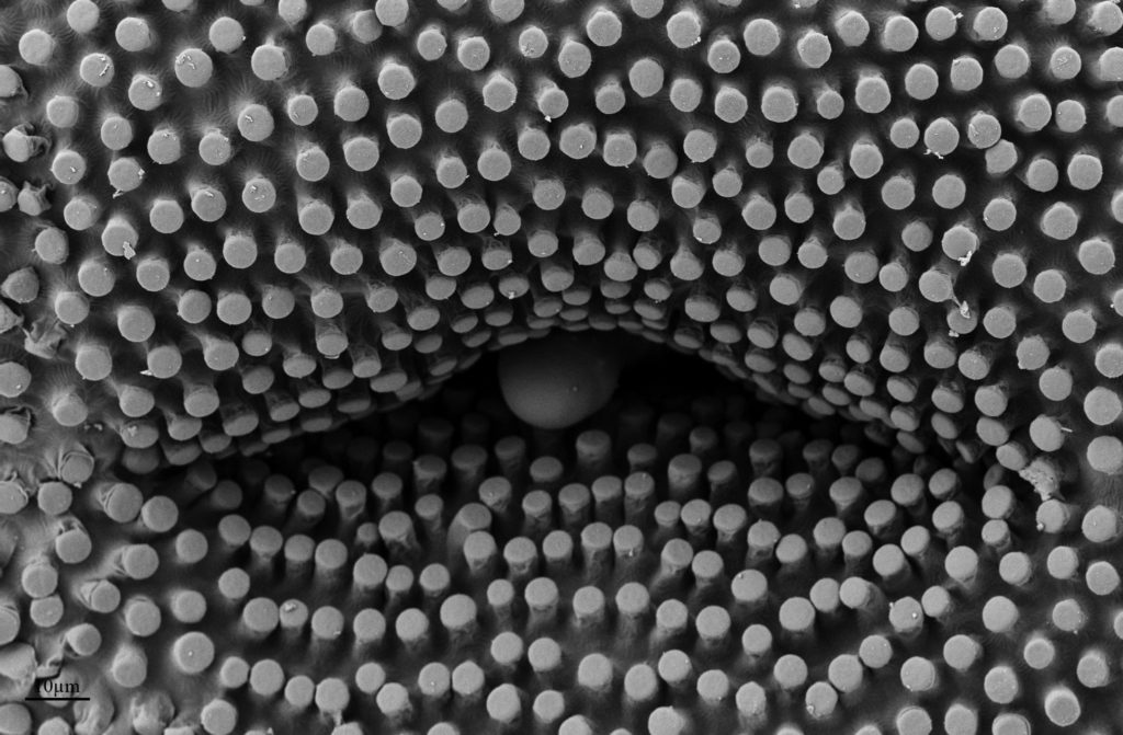
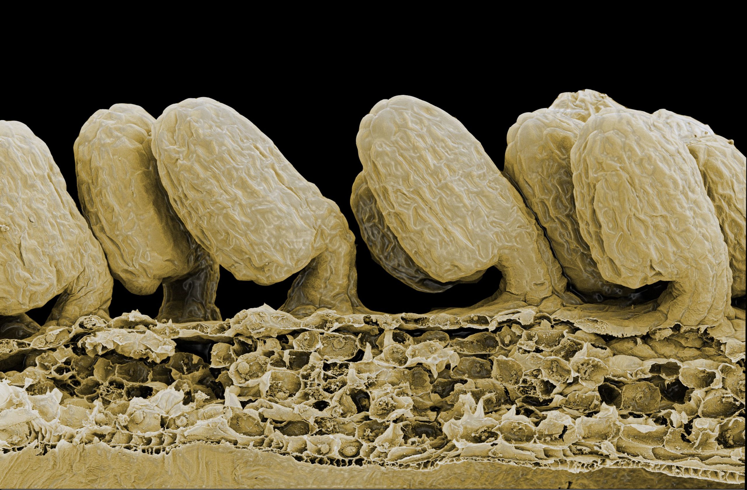
SEM image of the stigma surface of Streptocarpus suffruticosus (Gesneriaceae). 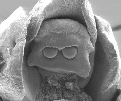
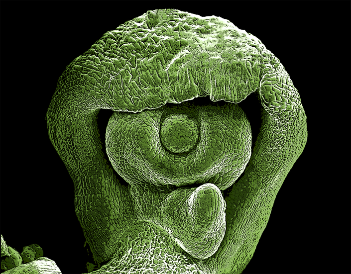
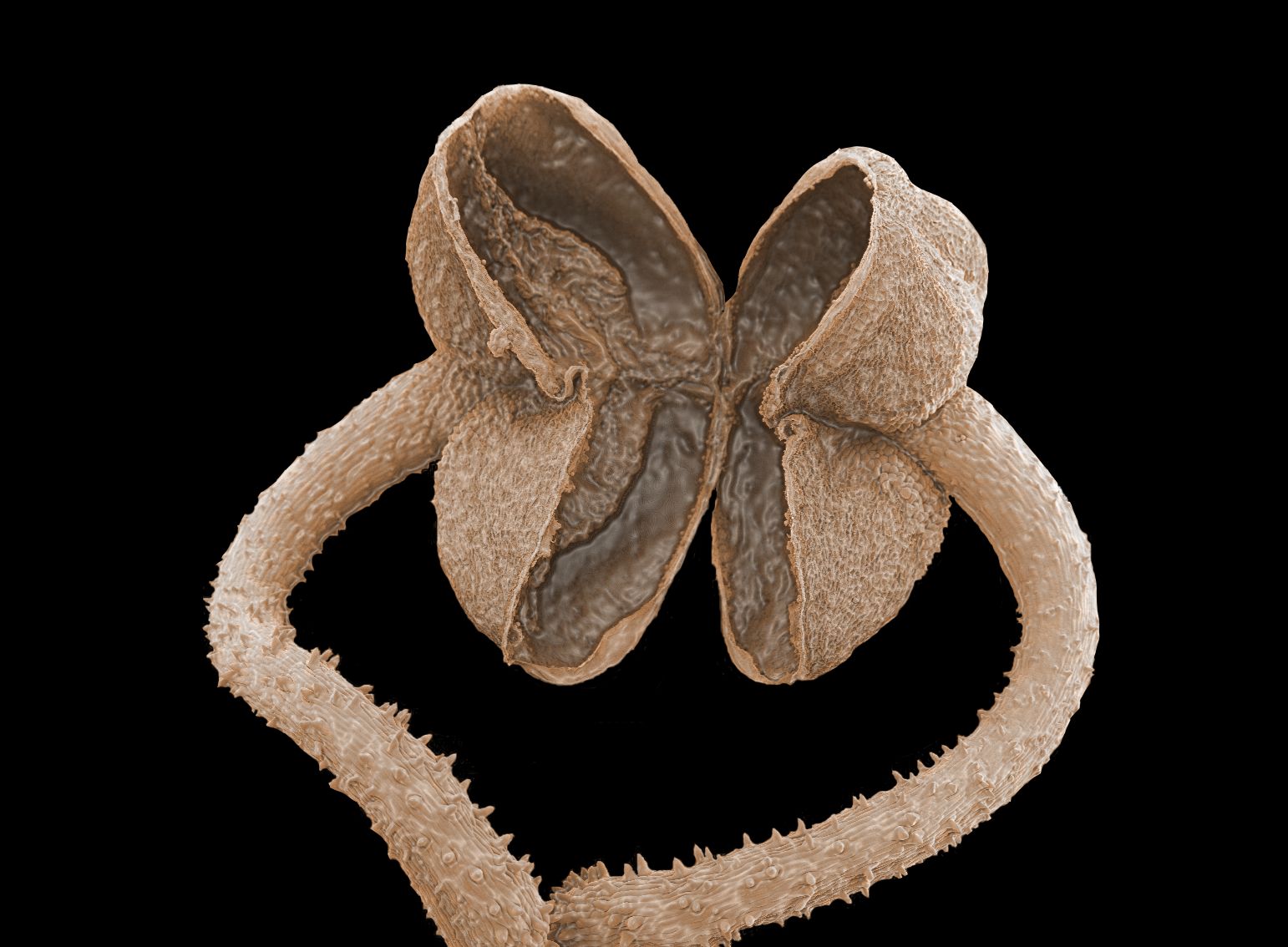
SEM of a receptive female cone with two nucelli developing in one epimatium of Acmopyle pancheri, projecting a ‘Darth Vader’-impression. 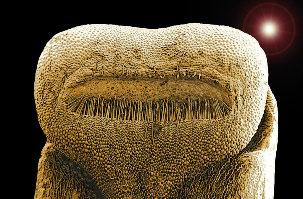
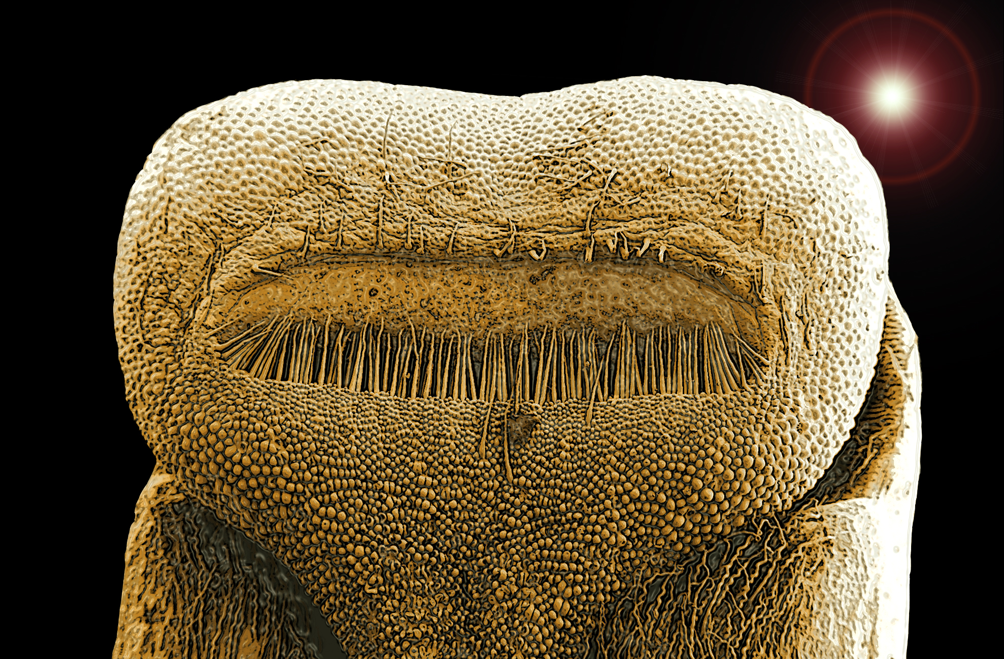
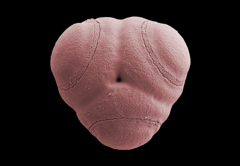
Etlingera albolutea stigma seen from below showing the ostiole bordered by a row of hairs. The ostiole is the receptive part where the pollen from the visiting pollinator is caught and starts growing a pollen tube along the 5.5 cm long style to ovary.