The following blog was written by Chris Knowles a digitiser in the Herbarium.
Since 2021 we have increased our digitisation capacity with the goal of getting to 1 million specimens imaged by Autumn 2024. Chris is databasing our fungal collections.
Part of my role involves checking the names and locations written on specimen packets, which means I get to see the huge range of habitats and substrates that the fungi have been collected from.
I wrote about a few of these in a previous blog: Towards 3 million: Fabulous fungi found in peculiar places – Botanics Stories, but couldn’t resist sharing some more that I have come across that have much grimmer origins … be warned.
As before, a great many specimens had been collected from within people’s homes and received at the RBGE for identification… with description notes such as ‘Growing out of house wall or windowsill or skirting board’ along with details of the house in question.
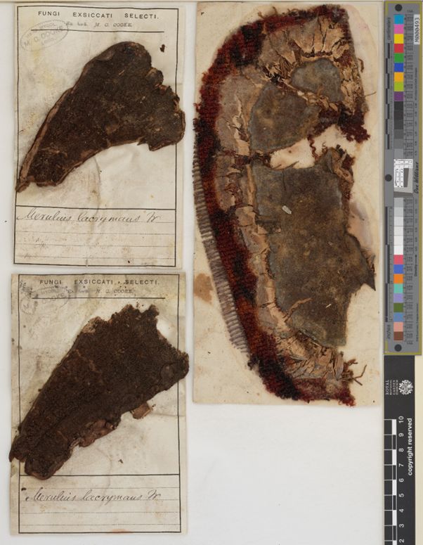
Nevertheless, one of the most feared species associated with houses is the Dry Rot fungus, (Serpula lacrymans) of which there are many specimens in the collection. Unfortunately, we now only have the record of a collection made from the “Free Church of Scotland, in cellars, growing on a bible”, that was collected in 1974. However, over a 100 years earlier the renowned mycologist Mordecai Cubitt Cooke collected these specimens on sections of carpet:
A little more surprisingly perhaps, has been the number of specimens associated with animals. Of course there are many that specialising in breaking down herbivore dung, but the Common Bird’s nest Fungus (Crucibulum laeve) described as growing on “rotting newspaper impregnated with goat urine” was a new one on me.
The collection here also has several examples of entomopathogenic fungi, those species that infect insects and modify their behaviour to improve spore dispersal. Tropical examples of this affecting ants were made famous by David Attenborough’s Planet Earth series, and also inspired the creation of the zombie mushrooms in the ‘The last of us’ games and television programme.
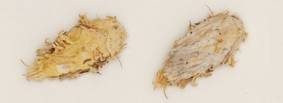
The image below is of a moth infected with Moth Urchin Fungus, the label of which states that it was found on a “Crag to west of Cnoc Daimh, Assynt. On carcass of a moth attached to Calluna” in 1996. While the specimen has preserved all the important macro and microscopic characters for identification, it has not quite retained its distinctive form, so I suggest an image search for ‘Akanthomyces aculeatus’ to see it in its full glory.
There are also specimens of Ascosphaera apis which causes something called Chalkbrood disease. Rather than altering an adult invertebrate’s behaviour, the spores of this fungus are accidentally fed to bee larvae in their cells within a hive, so the specimens in the collection are in tasty looking blocks of honeycomb. Once ingested the spores germinate and begin to draw nutrients from the bee larvae. Eventually the fungus grows out from the larvae, filling the entire cell with a chalky, white mycelium that mummifies the larvae before producing new spores to infect the food of other larvae within the hive.
Some other animal associated fungi in the collection came from mammals and fish, albeit dead ones. For example, we have a specimen of Chrysosporium pannorum, a fungus that breaks down the keratin that hair and nails are made of, that was collected from a “dead lamb carcass in derelict barn”. Topping that though, must be the specimen from the Cladosporium genus, which is a type of mould that was found “Growing on skull of blue finned tunny trawled up from 70-80 fathoms some 40 miles off Barra Head”, (Tunny was an older name for Tuna).
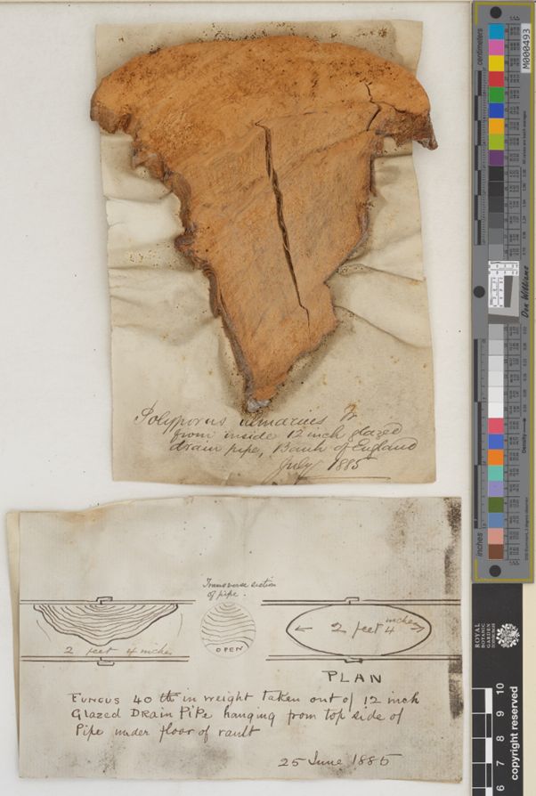
My favourite though, is the following specimen from a drainpipe that ran under the floor of a vault in the Bank of England, collected in 1885. I am particularly fond of the three-dimensional plan diagram that was drawn by the presumably incredulous collector. The large bracket-shaped fruitbodies of this fungus are mostly found on the trunks of broadleaf trees, (especially Elm) where they develop out from the fungal organism that will be feeding on the dead heartwood of the tree. The brackets are able to become so large because they are perennial, with a new, slightly broader fertile layer growing each year below the last. In this instance there must have been heavy wooden beams adjacent to the outside of the pipe joint, and the fine hyphal cells of the fungus found the space in the pipe to be the perfect place to spread and grow.